
SHEFFIELD, United Kingdom. A new ‘sat nav’ operating technique for surgeons could double the chance of survival of brain cancer patients by removing tumours which were thought to be inoperable.
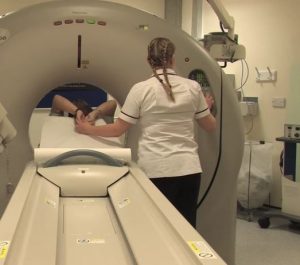
Sheffield Children’s Hospital has launched new surgical suite which includes an MRI scanner which can precisely monitor the brain during an operation.
It allows surgeons to work out whether they have managed to remove all of a brain tumour while still in theatre.
They can also work out precisely where a tumour is, thereby ensuring surrounding healthy tissue is not damaged.
Around 10,000 people are diagnosed with brain cancer each year and half will die. If a tumour is completely removed the chance of survival is up to 80 per cent. But if cancerous cells are left behind that falls to just 40 per cent.
Surgeons have already used the suite to carry out operations on two children and they hope to soon offer adults the benefit.
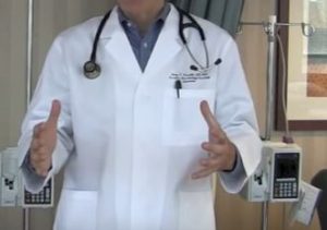
Hesham Zaki, head of the department of paediatric neurosurgery, said the equipment puts the Sheffield hospital at the forefront of increasing survival rates from brain tumours in the UK and worldwide.
He said: “The fact we can use the MRI scanner during the surgery is a real step-change. “Just like a sat nav, it tells me where I need to go.
“We scan the patient that we are operating on with their skull still open and the operation still ongoing.
“The MRI images mean that we can be sure the tumour has been completely removed and nothing has been left behind before we finish the operation.
“This is important because some types of brain tumour can look like normal brain. This is a sea-change. Tumours that were inoperable can now be operated on.”
Mr Zaki said children’s survival from brain tumours “is almost entirely dependent on whether the surgeon is able to remove all of the tumour”.
He said complete removal means there is a 70 per cent to 80 per cent chance of long-term survival.
“But if we leave some behind, this can drop to as low as 40 per cent,” he added.
Mr Zaki and colleagues use the MRI scanner in conjunction with “brain lab” technology, which enables them to pinpoint the exact location of a tumour in real time during surgery.
Surgeons use a medical probe or laser coming from the end of a microscope to “touch” tissue and tumour in the brain. The location of different tissue is then shown up on a screen which has been loaded with MRI images from the patient.
This enables surgeons to precisely work out where a tumour is, enabling it to be removed without harming healthy tissue.
Another MRI is then carried out during the operation to ensure all the tumour has gone before the surgery ends.
The hospital also has a new MRI suite for follow-up scans, which has removed the need for some children to go under general anaesthetic.
Jack McGuigan, an eight-year-old from Sheffield, has been attending Sheffield Children’s Hospital since he was 10 months old.
He has a condition called Langerhans’ cell histiocytosis (LCH), which is a cancer-like condition that causes growths of bone in his body.
Thanks to the scanner, he no longer needs general anaesthetic.
“It didn’t feel scary at all,” he said.
Ebony Taylor, 16, from Doncaster, has epilepsy and has also used the new scanner.
She said: “The old MRI scanner made such a horrible noise, I can’t describe it.
“I was nervous coming to use this one but it was so good. It wasn’t as claustrophobic and it was just more relaxing with all the lighting.”
Credit: The Telegraph (UK)